The Lateral Plantar Nerve

more nerves

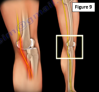
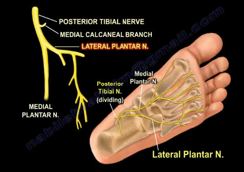
The lateral plantar nerve is branch of the posterior tibial nerve, which originates from the sciatic nerve. Around the medial side of the ankle, close to the tarsal tunnel, the posterior tibial nerve divides into the medial and lateral plantar nerves. anatomy

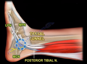
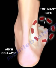
Thickening of the flexor retinaculum will cause compression of the posterior tibial nerve, which is called tarsal tunnel syndrome. When drawing a line between the medial malleolus and the calcaneus, the posterior tibial nerve divides into branches within 2 cm from this axis. The lateral plantar nerve is interesting because its branches give innervation to most of the intrinsic muscles in the foot—similar to the ulnar nerve in the hand. The lateral plantar nerve is also important due to its first branch being the Baxter’s nerve. baxter

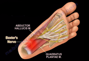
This nerve is always mentioned in nerve entrapment in running athletes and is associated with chronic heel related pain. The pain associated with the Baxter’s nerve is very similar to the pain associated with plantar fasciitis; the pain is in the same location, the mechanical symptoms are the same, and there is nerve pain unassociated with weight-bearing.

The first branch of the lateral plantar nerve gets compressed between the fascia of the abductor hallucis muscle and the medial side of the quadratus plantae muscle. This condition may require surgical release of the abductor hallucis fascia if conservative treatments and injections do not produce any effect.

In summary, the lateral plantar nerve:

  • Is similar to the ulnar nerve in the hand
  • Supplies most of the intrinsic muscles of the foot
  • Supplies the Baxter’s nerve branch
  • Can be injured during surgery (rod placement from the heel).

sustainAnother point of interest when it comes to the lateral plantar nerve is the hardware placement. Hardware placement prominent to the sustentaculum tali can injure the flexor hallucis longus tendon and the lateral plantar nerve.

Sever’s Disease

Sever’s disease is a common cause of heel pain in children between the ages of 9 and 12 years. The pain is due to calcaneal apophysitis occurring due to repetitive and continuous traction on the calcaneus from the Achilles tendon. The apophysis is not part of a joint and has muscle or tendon attachments. This traction apophysitis may lead to stress fractures, pain and tenderness over the heel.

Sever's1.png

Sever’s disease is similar to Osgood-schlatter disease of the tibial tubercle.

Sever's2.png

Patients are usually young athletes presenting with heel pain that increases with activities. Upon examination there could be swelling, tenderness, warmth and/or redness on the back of the heel where the Achilles tendon inserts.

Sever's3.JPG

Plain lateral X-rays may show sclerosis or fragmentation of the calcaneal tuberosity. Sclerosis is not specific for this condition.

Sever's4.png

Fragmentation of the calcaneal tuberosity on the other hand, is more common in patients with Sever’s disease relative to the general population.

Sever's5.png

Remember that Sever’s disease is a clinical diagnosis. X-rays may show other causes of pain such as tumors, fractures, infections or cysts. MRI is not commonly used, but can help rule out calcaneal stress fractures or osteomyelitis.

Sever’s disease is a self-limiting condition that usually resolves with time. Treatment usually consists of NSAID, Achilles tendon stretching exercises, and activity modifications and in severe condition a short leg walking cast can be used.

Gait

Gait is the pattern of how a person walks. We will be discussing different gait abnormalities.

Antalgic gait

Antalgic gait is a painful gait. A patient with antalgic gait does not want to spend time on the one leg due to pain. A patient wants to get their weight off the affected extremity. When pain is increased by walking, it leads to an antalgic gait (Figure 1).

gait1.PNG

An antalgic gait can be caused by multiple factors due to pain in any part of the lower extremity. It is usually caused from hip or knee pathology or from severe disc radiation symptoms (Figure 2).

gait2.PNG

The pain can be helped by using a cane on the opposite side of the painful extremity.

Trendelenburg gait

Trendelenburg gait is an abnormal gait that is usually found in people with weak abductor muscle of the hip which is supplied by the superior gluteal nerve. The patient cannot abduct the affected hip due weakness of the abductor muscles on the affected side. If the patient has weakness on one side of the pelvis and when the patient stands on that side, the pelvis on the contralateral side will drop. This is called Trendelenburg sign. A positive Trendelenburg sign occurs when there is dysfunction of the abductor muscles and the body is unable to maintain the center of gravity on the side of the stance leg (Figures 3, 4). The patient will show an excessive lateral lean to keep the center of the gravity over the stance leg.

gait3.PNG

gait4

Weakness can also occur in patients with L5 radiculopathy or avulsion of the abductor muscle tendon (Figure 5) which occurs with increasing frequency after hip replacement surgery.

gait5.PNG

The superior gluteal nerve injury is a major factyor in this gait. With bilateral weakness of the abductor muscles, the patient will have dropping of the pelvis on both sides during walking which leads to a waddling motion. This gait is seen in patients with myopathies.

Slap gait

Slap gait occurs due to weakness of the foot and ankle dorsiflexors which allows the foot slap down on the floor with each step. Slap gait is a heel gait abnormality that can be diagnosed by hearing the patient walk with a normal walking gait, the heel strikes the ground first followed by controlled relaxation of the foot and ankle dorsiflexors in order to allow the forefoot to come in contact with the ground

Steppage gait

Foot drop gait or steppage gait is due to total paralysis of the ankle and foot dorsiflexors (Figure 6). it is sometimes called neuropathic gait. A common symptom of foot drop is a high steppage gait that is often characterized by raising the thigh up in an exaggerated fashion while walking. The patient must externally rotate the leg or flex the hip or knee to raise the foot high enough to avoid dragging the toes along the ground. If the patient has foot drop then they have to have a high steppage gait or else they will trip on the foot and fall forward.

gait6.PNG

Conditions causing foot drop include L4-L5 disc herniation, a herniated disc compressing the L5 nerve root may cause foot drop, lumbosacral plexus injury due to pelvic fracture (Figure 7), hip dislocation leading to injury of the common peroneal nerve (Figure 8) and injury to the knee as knee dislocation (Figure 9).

gait7.PNG

gait8

gait9.PNG

Wide based gait

A wide based gait occurs due to myelopathy and neurological disorders. This gait disturbance is described as clumsy, staggering movements. It can be associated with cervical or thoracic spine pathology. Patient example of myelopathy with significant cervical spine disc compression of the spinal cord can be seen in Figure 10.

gait10.PNG

Patient will have a slow, wide, broad based ataxic gait. The patient will have a wide stance as they try to maintain balance. There will be unsteadiness of the trunk with excessive shift in the center of the gravity.

Gluteus maximus gait

When the gluteus maximus muscle (Figure 11) is week, the trunk lurches backwards (extension of the trunk). It occurs at heel strike on the weakened side to interrupt the forward motion of the trunk. This compensates for weakness of hip extension. The function of the gluteus maximus muscle is external rotation and extension of the hip joint.

gait11.PNG

Posterior Tibial Tendon Dysfunction

A tendon is a band of fibrous tissue that connects muscle to bone allowing the joint to bend. Tendons enable participation in physical activities such as running, jumping and other movements. The posterior tibial tendon starts in the calf and descends down the leg behind the inside of the ankle and attaches to the foot’s arch. Its function is to support the medial arch and sub-talar joint as the body passes over the foot. When the posterior tibial tendon becomes inflamed or is overstretched, the ability to support the arch is impaired resulting in flattening of the foot.

Posterior tibial tendon (Figure 1) dysfunction, as this phenomenon is called, can be attributed to several factors:

  • Tendon overuse.
  • exposing the foot to a significant load
  • Obesity
  • Hypertension
  • Trauma
  • Diabetes
  • Inflammatory diseases such as rheumatoid arthritis.

    post-tibial-tendon
    Figure 1

Patients with posterior tibial tendon dysfunction will often present with pain and swelling on the inside of the ankle, loss of the foot’s arch (flatfoot), tenderness over the mid-foot and an inability to stand on the toes.

To diagnose posterior tibial tendon dysfunction, physicians will likely use the “too many toes” test (Figure 2). Here, the physician measures abduction of the forefoot. If the

posterior-tibial
Figure 2

posterior tibial tendon is damaged, the forefoot will deviate outwards in relation to the rest of the foot and will appear to have too many toes when viewed from behind. In addition to the “too many toes” test, the physician may ask patients to do a single heel rise. Here, patients are asked to stand with their hands on the wall and lift the unaffected foot off the ground and raise the toes on the affected foot. If the heel does not rotate inward, there is posterior tibial tendon dysfunction.


Posterior tibial tendon dysfunction can be classified in four stages:

 STAGE I – characterized by an inflamed posterior tibial tendon with normal strength. Upon examination, the patient will be tender to palpation but may show little or no change in the arch of the foot. While X-rays will most likely show no changes, an MRI will likely reveal mild to moderate tenosynovitis.

 STAGE II – characterized by a partially torn tendon or degenerative changes. Here, the physician will note considerable flattening of the arch without arthritic changes and will have a positive too many toes sign. X-rays will reveal abduction of the forefoot while an MRI will reveal a partial tear.

 STAGE III – characterized by severe tendon degeneration with a rupture likely. Patients with stage III posterior tibial tendon dysfunction will present with rigid flatfoot. X-rays will likely reveal abduction of the forefoot and collapse of the talo-navicular joint while an MRI will show a tear in the tendon.

 STAGE IV – is similar to stage III with the addition of an arthritic ankle joint.

Treatment for posterior tibial tendon dysfunction can range from conservative to surgical depending on how far the condition has progressed. In its early stages, physicians will often utilize rest, anti-inflammatory medications, and immobilization. If the foot fails to respond to conservative treatment or the condition has progressed too far, there are several surgical procedures that can be utilized. First, physicians may perform a tenosynovectomy. Here, the surgeon will debride and excise inflamed tissue surrounding the tendon. A second option is an osteotomy. Here, the surgeon changes the alignment of the calcaneus and may remove a portion of the bone. A third option is a tendon transfer where fibers from another tendon are used to repair the posterior tibial tendon. Finally, surgeons may fuse one or more bone together, eliminating movement in the joint through a process called arthrodesis. During this procedure, the forefoot is stabilized.