The Ideal Patient With Low Back Pain

Low back pain is very common, and the majority of the patients get better with time. The ideal patient will get better with time and has no radiation below the knee, no history of trauma, no fever or chills or weight loss, no bladder or bowel dysfunction, no neurological deficits, and no pathological reflexes.

In order to optimize recovery, management of the patient should consist of early return to activity as tolerated, as the symptoms allow. You will give the patient reassurance with limited analgesia, early range of motion, and muscle relaxants. A healthy patient with an acute onset of non-traumatic low back pain, you do not need early diagnostic imaging before proceeding with the therapeutic treatment. Diagnostic imaging is not necessary unless the initial treatment is unsuccessful, and the symptoms are prolonged. X-rays may not be needed in the first six weeks unless there is a reason for it, such as red flags. In fact, the use of x-rays can lead to better patient satisfaction but doesn’t necessarily lead to better patient outcome. X-rays and MRIs may show changes in the intervertebral discs and may be associated with the patient’s pain, but these changes are also commonly seen in cross-sectional studies of asymptomatic people. There are a lot of false positive MRIs, and you need to correlate the MRI findings with the clinical findings. Don’t rely on the MRI alone! Just because you have MRI changes or disc protrusion, it does not mean that the patient needs surgery!

A nonspecific pain does not require surgery; therefore, it does not require further work-up. There are risk factors associated with low back pain that includes Poor physical fitness; Smoking; History of repetitive bending or stooping on the job and whole-body vibration exposure. If the patient has a simple low back pain, 50% of the patients resolve their pain in one week. Resolution of the acute back pain occurs in 90% of the patients within one month. If the patient has leg pain greater than back pain, then the patient has sciatica. Sciatica means nerve root irritation, probably due to a herniated disc.
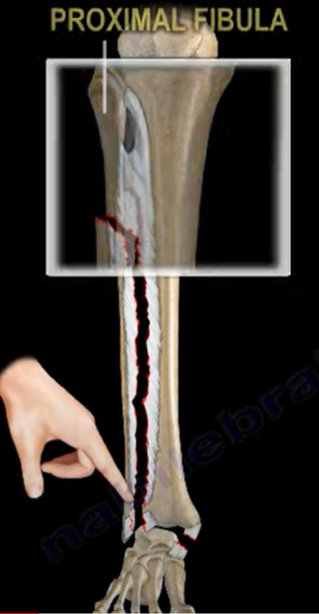
Ankle fracture Maisonneuve Fracture – Everything You Need to Know

Maisonneuve fracture involves fracture of the proximal fibula associated with an occult and unstable injury of the ankle. The problem in these patients occur when the ankle injury is presented without a fracture of the lateral malleolus, or the medial malleolus and the injury is mistakenly diagnosed as an ankle sprain and the proximal fibular fracture is missed. Examine the leg for tenderness in the proximal fibula to diagnose a proximal fibula fracture. The patient could be mistakenly treated for having an isolated proximal fibular fracture alone and the ankle injury is missed.

 High index of suspicion is necessary to diagnose and treat this injury. Maisonneuve fracture equals syndesmotic injury. Syndesmotic Injury equals Syndesmotic Reduction and Fixation. If ankle x-rays show medial or posterior malleolus fracture, or a medial clear space widening with no fracture of the lateral malleolus, then you must obtain a long-leg films to assess possible proximal fibular fracture. Clinical examination of their entire leg for pain and tenderness in addition to long leg films of the entire leg that includes the ankle, and the knee is mandatory in case of the patient with approximate fibular fracture to exclude the presence of an additional ankle injury, or if the patient has an unexplained increase in the medial clear space of the ankle joint. You should be searching for the presence of a high fibular fracture. Look for signs of syndesmotic injury such as an unexplained increase in medial clear space or tibiofibular clear space is widened and it should be less than 5 millimeters.

So how do you explain this injury? It is explained by the presence of rotation force to the ankle with transmission of the force through the interosseous membrane, which exits through a proximal fibular fracture. Maisonneuve fracture occurs from external rotation of the foot, most often with pronation mechanism. This force has to go somewhere! If you don’t see a fracture of the fibula then do the squeeze test or the external rotation stress test (both will show syndesmotic). The injury can involve the deltoid ligament injury or medial malleolar fracture medially and a fibular fracture proximally. Additionally, the tibiofibular ligaments are also involved, which can be the anterior tibiofibular ligament, interosseous ligament, the posterior tibiofibular ligament or posterior malleolar fracture. This looks like a very unstable ankle injury that may not be very obvious at presentation and you have to look out for it.

So how do you treat an Maisonneuve Fracture? This treated by fixation of the tibiofibular syndesmotic injury (key of treatment) or syndesmotic screws. if you have a medial site injury and there is a tear of the deltoid ligament, leave it alone. if there’s a medial malleolus fracture you should fix that of the lateral side if there’s approximate fibular fracture leave it alone. If there is a medial malleolar fracture, it should be fixed. If there is a proximal fibular fracture on the lateral side, leave it alone. As for the Syndesmotic Injury, the fixation has to be stable and adequate. Because of the magnitude of the injury, the Maisonneuve fracture may require more syndesmotic screws than with a routine ankle fracture with syndesmotic injury. After the fixation you will give a short leg non-weight bearing splint for six to eight weeks. Here is a patient taste example: the proximal fibular fracture and you can see increase in the medial clear space and you can see that the syndesmosis is widened. You can see that in the posterior malleolar fracture the patient is fixed with syndesmotic screws.

The Ideal Patient with Low Back Pain – Everything You Need to Know

The ideal Patient with Low Back Pain – Everything you need to know

Low back pain is very common, and the majority of the patients get better with time. The ideal patient will get better with time and has no radiation below the knee, no history of trauma, no fever or chills or weight loss, no bladder or bowel dysfunction, no neurological deficits, and no pathological reflexes.

In order to optimize recovery, management of the patient should consist of early return to activity as tolerated, as the symptoms allow. You will give the patient reassurance with limited analgesia, early range of motion, and muscle relaxants. A healthy patient with an acute onset of non-traumatic low back pain, you do not need early diagnostic imaging before proceeding with the therapeutic treatment. Diagnostic imaging is not necessary unless the initial treatment is unsuccessful, and the symptoms are prolonged. X-rays may not be needed in the first six weeks unless there is a reason for it, such as red flags. In fact, the use of x-rays can lead to better patient satisfaction but doesn’t necessarily lead to better patient outcome. X-rays and MRIs may show changes in the intervertebral discs and may be associated with the patient’s pain, but these changes are also commonly seen in cross-sectional studies of asymptomatic people. There are a lot of false positive MRIs, and you need to correlate the MRI findings with the clinical findings. Don’t rely on the MRI alone! Just because you have MRI changes or disc protrusion, it does not mean that the patient needs surgery!

A nonspecific pain does not require surgery; therefore, it does not require further work-up. There are risk factors associated with low back pain that includes Poor physical fitness; Smoking; History of repetitive bending or stooping on the job and whole-body vibration exposure. If the patient has a simple low back pain, 50% of the patients resolve their pain in one week. Resolution of the acute back pain occurs in 90% of the patients within one month. If the patient has leg pain greater than back pain, then the patient has sciatica. Sciatica means nerve root irritation, probably due to a herniated disc.

Frozen Shoulder – Adhesive Capsulitis

Frozen Shoulder Adhesive Capsulitis – Everything You Need To Know

Frozen shoulder (adhesive capsulitis)

The frozen shoulder can be associated with diabetes or thyroid disease.  It may be the initial presenting symptom for these conditions.  The exact details of this relationship remain poorly understood.  Ever patient with a frozen shoulder should have the HbA1c and TSH levels tested.  Also, check for arthritis, rheumatoid factors and antinuclear antibodies.  Most patients with frozen shoulder are female between the ages of 40 and 60 years old.  And frozen shoulder, the patient will lose both the active and the passive range of motion of the shoulder.  The patient develops pain, which means that there is inflammation with early fibrosis of the joint capsule, leading to joint stiffness.  The active and passive global motion, especially external rotation, will be reduced compared to the other side.  The shoulder pain and motion loss is usually not related to trauma.  It is an idiopathic process that results in shoulder pain and loss of motion due to contracture of the capsule.  The essential lesion involves the coracohumeral ligament and the rotator interval.  The synovial inflammation and capsular fibrosis results in pain and joint volume loss.  Check for previous trauma or fractures.  Rule out shoulder joint arthritis and rule out posterior dislocation of the shoulder.  It can also occur post-surgery from a rotator cuff tear.  It may also be associated with dupuytren disease and cervical disc disease.  Check for medical comorbidities such as stroke or cardiac diseases.  The x-ray will exclude trauma, malignancy, arthritis, calcific tendinitis, impingement, and AC joint arthritis.  And frozen shoulder, the humeral head will remain in its normal location.

MRI

There is a space reduction in the axillary recess.  Rotator cuff strength is normal by exam and on the MRI.  The pain and stiffness lasts beyond 6 months, then you can do manipulation of the shoulder under anesthesia.  There is a 50% failure rate in diabetics.  The diabetes is associated with a much worse prognosis and poor outcome for surgical and nonsurgical treatment.

Treatment

·         Nonoperative

o   This should be done for at least 3 to 6 months.

o   Supervised or home-based capsular stretching program +/-intra-articular steroid joint injection.

o   Nonsteroidal anti-inflammatory medication

·         Surgery

o   Capsular release (arthroscopic or open) and release the intra-articular and subacromial adhesions.

o   The axillary nerve may be injured during release of the capsule.

o   Utilize surgery in patients that have failure of initial conservative treatment for 3 months, and the patient remains functionally limited.