Traumatic Neonatal Distal Femoral Physeal Injury

physeal

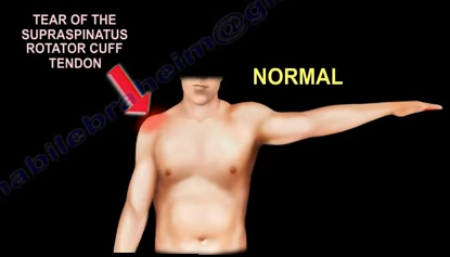
A physeal injury in the distal femur in the neonate is rare. In general, traumatic neonatal physeal fracture usually occurs in the distal humerus and rarely occurs in the distal femur. The condition usually occurs due to birth trauma and usually results due to physeal separation which results in epiphyseal separation. The thigh will be swollen and there may be hypomobility of the extremity. The neonate will be fussy or irritated with significant swelling in the thigh. The condition is under diagnosed. The epiphysis is usually present at birth on x-rays. This condition can become complicated if the epiphysis is not completely ossified at birth or if the child is born prematurely. In this situation, the x-ray interpretation may be difficult. An MRI will be really helpful if the doctor is uncertain of the diagnosis. X-rays should be taken at the child at birth. The physician should look at the lateral x-ray and find the epiphysis is present and ossified at birth with varying degrees (may not be clear). Each epiphysis will line up with its corresponding bone. Epiphysis of the distal femur should line up with the femoral congenital kneeshaft. Epiphysis of the proximal tibia should line up with the tibial shaft. If the two epiphysis separate from each other, then this is a congenital dislocation of the knee. This can be a spectrum of injury that varies from hyperextension to subluxation, to frank dislocation. If the epiphysis is separated from its corresponding bone, for example, the epiphysis of the distal femur is separated from the shaft of the femur, then this is a physeal injury. Because the distal femur is mostly cartilaginous, you don’t see bone shifting, you see a little hint of the separation of the physis by seeing that the small ossified epiphysis is not in its normal position that corresponds with the axis of the bone. You need to know the difference between physeal injury and congenital dislocation of the knee. Accurate, gentle closed reduction with follow-up x-rays to confirm the reduction and to detect any early bony bridging.


Drop Arm Test

The Drop Arm Test is used for diagnosing rotator cuff tendon tears, specifically the supraspinatus tendon tear. The test helps in determining if there is a tear in the supraspinatus tendon and the can be done by many techniques. The patient should be instructed to fully abduct the arm. Then, the patient should slowly lower the arm to the side. If there is a tear in the rotator cuff tendon, the arm will drop from a position of about 90° of abduction. tearEven if the patient attempts this several times, the patient with a supraspinatus tendon tear cannot lower the arm smoothly and slowly. If the patient with the supraspinatus tendon tear is able to hold the arm in abduction, a small amount of pressure on the forearm will cause the arm to fall to the side.

Another method of performing the test begins with the physician instructing the patient to fully abduct the arm and the examiner supporting the arm so that it is able to be tested in 90° of abduction. The patient is asked to actively lower the arm from abduction to the side, slowly andtendon tear smoothly in a controlled way. A positive test occurs when the patient is unable to hold the arm in 90° of abduction or is unable to control lowering the arm to the side. The practical way of performing the test is when the examiner holds the arm fully abducted by the side of the patient and then will release it. If the arm drops, this will mean that there is a large rotator cuff tear present. The test is positive when pain and weakness causes the arm to drop to the side.


Spine Concepts- Cervical Rheumatoid Arthritis

Cervical spine involvement occurs in about 90% of patients with rheumatoid arthritis. All rheumatoid arthritis patients should have a cervical spine examination. The physician should begin with getting cervical spine x-rays because this helps to diagnose atlantoaxial instability. Early aggressive medical treatment can decrease this risk. C1-C2 instability is common and can occur in up to 80% of cases. C1-C2 instability occurs due to transverse ligament pathology. Flexion/extension views will be beneficial for patients with rheumatoid arthritis, especially preoperative x-rays. If it looks bad, stabilize the spine before performing total hip or total knee procedures. Discover the C1-C2 instability and fix it first adibefore doing elective total hip procedure. The normal A.D.I. in adults is less than 3mm. A.D.I. more than 3.5 mm indicates instability of the upper cervical spine may be present. A.D.I. more than 7mm indicates disruption of the alar ligament and these patients can have cervical spine myelopathy. The Atlanto-Dental Interval (A.D.I.) is an unreliable predictor of paralysis. The posterior atlanto-dental interval is a better screening test that can predict spinal cord injury. If the Posterior Atlanto-Dental Intreval (P.A.D.I.) is less than 14mm, this can predict a spinal cord injury. An MRI should be obtained. Surgery is performed if the A.D.I. is more than 10mm or if the P.A.D.I. is less than 14mm. Surgery is done by C1-C2 fusion.

Clinically, the C1-C2 instability could give neck pain, headache, and myelopathy with abnormal gait, paresthesia, and difficulty in fine motor control. Basilar Invagination occurs in about 40% of patients with rheumatoid arthritis. Basilar impression (invagination) occurs if the odontoid process tip is 5mm or more above Chamberlain’s line. In this case, do occiput to C2 fusion, plus or minus odontoid resection. Subaxial subluxation occurs in about 20% of patients and the indication for surgery is neurological compromise. If the space available for the spinal cord is less than 14mm, then do surgery (posterior fusion). Surgery is usually not successful with severe types of neurologic impairment.

extensionSurgery should be performed if the patient has severe pain, neurological deficit, and x-rays showing that the P.A.D.I. is less than 14mm. Surgery should also be performed in cases of superior odontoid migration and subaxial subluxation with the sagittal canal diameter being less than 14mm. If the posterior atlanto-dental interval is more than 14mm, the patient will demonstrate significant motor recovery after surgery.

spinal cord compression