The motions of the thumb are complex and are often difficult to visualize, as multiple joints and planes are involved. The motions are crucial to the overall function of the hand, with amputation of the thumb resulting in 40% impairment. Amputation has also been cited as causing 22% disability of the whole person. In cases of thumb hypoplasia and carpometacarpal joint instability, thumb amputation and index finger pollicization is recommended. Because of the value of the thumb, reimplantation of the amputated thumb at any level is recommended. The main motions of the thumb include flexion, extension, abduction, adduction, and opposition. Valued as 50-60 percent of overall thumb function, opposition is one of the most critical motions. Abduction and adduction of the thumb occur mainly at the carpometacarpal (CMC) joint. Abduction is described as the motion of the thumb away from the second metacarpal, whereas adduction is the opposite of the motion. These motions are defined in two planes: palmar and dorsal. In radial abduction-adduction, the thumb moves parallel to the radius and the palm of the hand. A smaller range of motion is generally seen in the radial plane. Alternatively, in palmar abduction-adduction the thumb follows a plane perpendicular to the palm of the hand. Clinically, these motions are not typically measured directly but are assessed in the motion of opposition. Flexion and extension can occur at all three joints of the thumb, including the CMC, MCP, and IP joints. These motions at the CMC joint are often not measured, as they are challenging to quantify. The average flexion observed at the MCP joint is approximately 50 degrees, although this may be limited in some individuals to less than 30 degrees. Extension is not usually seen at the MCP joint. Flexion at the IP joint approaches 80 degrees on average. Typically, the IP joint is capable of approximately five degrees of extension. Opposition is a very important motion and, as mentioned, 50-60% of overall thumb function. It is comprised of several integrated motions: palmar abduction that transitions to adduction at the CMC joint, rotation at the CMC joint, and flexion at the CMC, MCP, and IP joints. The composite motion involves crossing the thumb over the thumb of the hand toward the base of the little finger. Complete opposition of the thumb may be defined in two ways: by the tip of the thumb touching the base of the fifth digit and by the tip of the thumb touching the tip of the little finger. Therefore, deficiencies in opposition can be quantified by measuring the distance from the tip of the thumb to the base of the little finger or from the tip of the thumb to the tip of the little finger. Alternatively, the degree of opposition may be determined by measuring from the flexor crease of the IP joint to the distal palmar crease over the third metacarpal. A normal span is at least 8 cm. opposition may also be assessed by systematically touching the tip of the thumb to various parts of the hand, a clinical test outlined by Kapandji.
Hip dislocation can be either a simple dislocation or it can be a fracture dislocation which involves the posterior wall of the acetabulum or the femoral head. Dislocation of the hip can be two types: posterior dislocation (most common type) or anterior dislocation (rare). Position of the hip during the impact decides the injury. In posterior dislocation of the hip, which is the commonest type, the lower limb will be flexed, adducted, and internally rotated. Anterior hip dislocation is rare. It could be a superior anterior hip dislocation. The limb will be extended, abducted, and externally rotated. With an anterior inferior dislocation (obturator type), the extremity will be flexed, abducted, and externally rotated. Hip fractures are different than hip dislocations. Notice that the affected extremity is shortened and externally rotated with a hip fracture. Hip dislocation of any type is an emergency. It must be reduced in less than 6 hours of injury. After reduction of the hip, get a CT scan. Although x-ray is helpful, a CT scan clearly outlines the bony injury. Check the CT scan for congruous reduction, for absence of fracture, and absence of marginal impaction in the acetabulum (with posterior wall fracture, check for marginal impaction). Marginal impaction is more common in posterior acetabular wall fractures and could lead to instability. Displaced or comminuted posterior wall fracture could lead to arthritis. Make sure that you have good congruous reduction with no loose bodies or important fractures present. Check for fractures of the acetabulum and the size of the fragment. The size of the posterior wall fracture has an effect on the stability of the hip joint. If congruous reduction of the hip is not obtained, perform open reduction urgently. Open reduction can be done through an anterior approach or a posterior approach. Hip dislocation with or without associated fracture can cause complications. The risk of avascular necrosis depends on the interval between the injury and reduction of the dislocation. Urgent reduction of hip dislocation is mandatory to avoid AVN and interruption of the blood supply which leads to collapse of the femoral head. Reduce the hip and recheck the sciatic nerve function. Always reduce the hip early. Closed reduction should be done in less than 6 hours. When injury occurs to the sciatic nerve due to posterior hip dislocation, the common peroneal nerve is usually affected, causing weakness in dorsiflexion of the ankle and loss of toe extension. Injury can occur in varying degrees of severity and it can be missed. Check for foot drop. Movement of the toes may be misleading. Movement of the toes may appear as dorsiflexion, however this really is the result of plantar flexion. Documenting the injury is important to avoid medical legal problems. Injury to the sciatic nerve usually occurs from the dislocation and not from the reduction of the hip. The longer the wait for the reduction of the dislocation, the more the patient is predisposed to sciatic nerve injury. The length of time a hip remains dislocated influences the incidence and the severity of a major sciatic nerve injury. Patient recovery of the sciatic nerve occurs in 60-70% of patients. The patient usually requires an anti-foot drop splint to prevent equinus of the ankle. There is approximately 10% incidence of sciatic nerve palsy from posterior hip dislocation. Neurologic examination at the time of injury is usually difficult, however, it is extremely necessary. Check for sensation on the top of the foot. In posterior dislocation of the hip, always look for injuries in the knee such as with a dashboard injury. The force of the injury is usually transmitted from the knee to the hip. There may be an associated posterior cruciate ligament (PCL) injury or a meniscal tear. Examine the knee for injuries and an MRI of the knee may be needed. In cases of high energy trauma, always look at the chest. There might be a tear of the aorta. Check for widening of the mediastinum on chest x-rays. There is concern of deceleration injury involving the aorta. You may apply advanced trauma life support (ATLS) protocol. More flexion, internal rotation, and adduction favors pure dislocation of the hip. Less flexion, internal rotation, and adduction favors fracture dislocation of the hip. Hip dislocation may be associated with acetabular fracture or fracture of the femoral head (Pipkin fracture). With Pipkin fracture, as the femoral head dislocates, it hits the posterior wall of the acetabulum and the femoral head fractures. This may be different from an anterior hip dislocation. Anterior hip dislocation will cause impaction of the femoral head or indentation fractures. Classically, Pipkin fracture is a posterior dislocation of the hip and fracture of the femoral head. To treat this, do emergency closed reduction of the hip within 6 hours. Closed reduction is done to avoid avascular necrosis (AVN) of the hip. Reduction of the hip joint and mobilization of the patient with protected weight-bearing crutches for 4-6 weeks. After closed reduction, when the patient has an associated fracture, assess the hip stability, especially if the fragment is not too large. The hip is usually stable if the fragment size of the acetabulum is less than 20%. More than 40%, the hip is unstable. Between 20%-40% fragment size, the hip stability is undetermined. When there is an associated acetabular fracture, the best method to assess the stability of the hip is by examination of the patient under general anesthesia utilizing fluoroscopy. Asses the posterior wall stability with the obturator oblique view. Hip will be in flexion, adduction, and add axial load. Check the medial clear space for opening (opening of the medial clear space suggests instability of the posterior wall fracture. Irreducible isolated posterior dislocation; do emergency surgical treatment to reduce the hip. If there is an associated acetabular fracture or femoral head fracture, do urgent closed reduction of the hip dislocation followed by stabilization of either of the fractures if needed according to the protocol. For a posterior hip dislocation with posterior acetabular wall fracture, you must assess the stability of the hip joint by examination under anesthesia after closed reduction. After closed reduction, if the dislocation is not congruent, do open reduction and fixation urgently. For a Pipkin femoral head fracture, do headless screw fixation.
Pain, numbness, and paresthesia in the palmar aspect of the thumb, index, and long finger (median nerve distribution). Symptoms of carpal tunnel syndrome occur more at night. These symptoms wake the patient up from sleep, causing the patient to shake the hand in attempt to resolve these symptoms. Percussion of the volar wrist crease produces electric sensation distally to the fingers. Phalen’s maneuver is performed by flexing the wrist for 60 seconds. This will increase the carpal tunnel pressure temporarily and produce the symptoms. If the test is positive, the patient will have numbness and tingling in the hand and wrist. The Positive Compression Test (Durkan’s Test) is the most sensitive test. The examiner places even pressure with two thumbs directly over the patients median nerve in the carpal tunnel for about 30 seconds. Reproduction of symptoms in the distribution of the median nerve means that the test is positive for carpal tunnel syndrome. Self-administered hand diagram is extremely helpful (most specific test for carpal tunnel syndrome). The patient should highlight the areas where they are experiencing the symptoms. The patient may complain of thenar atrophy, weakness, or clumsiness of the hand. The patient’s history and examination is an indication for carpal tunnel syndrome. Carpal tunnel syndrome is a clinical diagnosis. Carpal tunnel syndrome can be treated by anti-inflammatory medication or activity modification. Activity modification includes avoiding activities that aggravate the symptoms. It can also be treated with neutral wrist splints. It can help night time symptoms because it lowers the carpal tunnel pressure. Functional wrist splints (30o extension) will aggravate carpal tunnel syndrome because it increases the carpal tunnel pressure. At 3 months, 50% of patients will improve with splints. At 18 months, more patients will improve with splints. Sometimes, I use Vitamin B6. There is really no proof that vitamin B6 and physical therapy have any significant effect on improvement of carpal tunnel symptoms. Steroid injections are used for the treatment and for diagnosis of carpal tunnel syndrome if clinical examination or electro-diagnostic test is not clear. If the patient temporarily improves from injection, then the patient will definitely improve from surgery. For steroid injection, mark the intersection of the palmaris longus tendon and the distal palmar crease. Next, go 1 cm proximal and 1 cm ulnar to that site, this will be the point of injection. Use a 25 gauge needle with desired steroid and 1 mL of 1% lidocaine. Put the needle at a 45o to the skin of the wrist. Direct the needle towards the base of the thumb and advance the needle distally and slowly. The physician should warn the patient before the injection that if any feeling of numbness, paresthesia, or severe pain exists to let the physician know about it. Injection gives 80% transient improvement and 22% of the patients will be symptom free 1 year after injection. Carpal tunnel release surgery can be open or endoscopic. Surgery is usually done when there is persistence of the symptoms and failure of nonoperative treatment. The injection is a good prognosis for improvement after surgery when the splint no longer works, and when steroid injection only gives temporary improvement (injection is a good prognosis for improvement from surgery). The median nerve is much like a truck passing through a tunnel. The truck (nerve) should be able to pass through the tunnel with ease and without friction. If the tunnel is narrow then the nerve (truck) cannot pass. If you want the nerve to pass, then widen the tunnel. The tunnel is widened by cutting the transverse carpal ligament, as seen in this example. The American Academy of Orthopedic Surgeons (AAOS) recommends doing electrodiagnostic studies before performing carpal tunnel release surgery. Graham stated, that if the patient has a strong history and clinical examination for the diagnosis of carpal tunnel syndrome, then the electrodiagnostic test is unlikely to change the clinical diagnosis. Endoscopic procedure will give a better early rehab. The result is the same as with an open release, however incomplete release is a complication of the endoscopic procedure. The pinch strength returns to normal by 6 weeks. The grip strength returns to normal by 12 weeks. At one year, 20% of patients with severe carpal tunnel symptoms will continue to have symptoms. Revision carpal tunnel usually occurs when there is incomplete release. 25% will have no relief. Only 25% will have complete relief. The recurrent motor branch of the median nerve can be injured during the surgery. I want to talk about the anatomy and the variation in distribution of this nerve. After passing through the carpal tunnel, the median nerve gives a branch on the radial side called the recurrent motor branch. The recurrent motor branch is an important nerve supply to the thenar muscles. The recurrent motor branch of the median nerve has multiple variations of the nerve: 30% are subligamentous with recurrent innervation and 20% are transligamentous with recurrent innervation. If this nerve is injured, the patient will get progressive thenar atrophy due to that injury. It is important to cut the transverse carpal ligament far ulnarly to avoid cutting the recurrent motor branch of the median nerve. If you see a patient after carpal tunnel release and that patient has progressive thenar atrophy, this can be explained by the fact that there is an injury to an unrecognized transligamentous motor branch of the median nerve.
Girdlestone Procedure for Femoral Neck Fractures in the Elderly
Girdlestone procedure is a salvage procedure. It means removal or resection of the neck of the femur. The diseased femoral head is cut off with a bone saw. It means removal or resection of the head and neck of the femur. The affected femoral head is removed as you can see in this picture. Girdlestone is usually done in the following situations: patient has a severely painful hip and a total hip replacement cannot be done such as in cases of severe infection of the hip or in a nonabulatory cerebropalsy patient with a painful hip dislocation. It can also be done in selective tumors of this area. The procedure is referred to as a “salvage” procedure. It is the lesser of two evils or it is the final alternative procedure. This procedure may have a role in cases of displaced femoral neck fractures or in cases of failed internal fixation of femoral neck fractures in debilitated elderly patients. Let’s agree that in the elderly patient, hip prosthesis either unipolar or bipolar and usually cemented, is the ideal surgical procedure for displaced femoral neck fractures, especially if the patient is debilitated and old. In an active elderly patient, total hip replacement should be considered. Sometimes the medical condition and the age of the patient does not support or allow the use of a prosthesis in the elderly. I use the girdlestone procedure in some cases of displaced femoral neck fractures in the elderly, especially if the patient is debilitated and nonambulatory, and when the medical comorbidities are almost prohibitive for surgery. Comorbidities include chronic renal failure, COPD, and congestive heart failure. Even if the prosthesis could be done, the pre-injury cognitive and physical function is predictive of post-operative functional outcome after hip fracture surgery and this select group of patients will not be functional with the prosthesis. The purpose of the Girdlestone procedure is to decrease the pain and to preserve the life of the patient despite the considerable shortening of the extremity. It is an alternative to hospice or alternative care. It is the simplest and the least complex procedure for the patient. Counseling to the patient and the patient’s family should be done. The Girdlestone procedure can be done anteriorly or posteriorly. You do not need traction post-operatively. You should get the patient out of bed immediately, and you should do physical therapy early. You will keep the patient in a step down or ICU for a few days after surgery. The patient should be admitted by the geriatric services in cooperation with the trauma services. Surgery should be done within 48 hours or as soon as the patient is optimized medically because that could decrease the mortality rate. Sometimes optimization of the patient is not that easy. The mortality rate is 25% at one year and 6% during hospitalization. The pre-injury mobility is the most significant determining factor for post-operative survival. In patients with femoral neck fractures, surgery done on weekends was associated with an increase in hospital mortality rate, so it is better to do this surgery on week days. If you try to do a simple procedure such as fixation of the displaced femoral neck fracture, the failure rate is about 46% with fixation techniques in the elderly. There is a growing number of people over 90 years of age who will suffer from femoral neck fractures and these patients will need decisions and appropriate care for their situation. Advanced age is associated with increased mortality and poor functional recovery, so we need to think of new ways to approach the increased number of femoral neck fractures in the elderly, and I think that Girdlestone procuedre should be utilized in some select indications.
 Because of the value of the thumb, reimplantation of the amputated thumb at any level is recommended. The main motions of the thumb include flexion, extension, abduction, adduction, and opposition. Valued as 50-60 percent of overall thumb function, opposition is one of the most critical motions. Abduction and adduction of the thumb occur mainly at the carpometacarpal (CMC) joint. Abduction is described as the motion of the thumb away from the second metacarpal, whereas adduction is the opposite of the motion. These motions are defined in two planes: palmar and dorsal. In radial abduction-adduction, the thumb moves parallel to the radius and the palm of the hand. A smaller range of motion is generally seen in the radial plane. Alternatively, in palmar abduction-adduction the thumb follows a plane perpendicular to the palm of the hand. Clinically, these motions are not typically measured directly but are assessed in the motion of opposition. Flexion and extension can occur at all three joints of the thumb, including the CMC, MCP, and IP joints. These motions at the CMC joint are often not measured, as they are challenging to quantify. The average flexion observed at the MCP joint is approximately 50 degrees, although this may be limited in some individuals to less than 30 degrees. Extension is not usually seen at the MCP joint. Flexion at the IP joint approaches 80 degrees on average. Typically, the IP joint is capable of approximately five degrees of extension. Opposition is a very important motion and, as mentioned, 50-60% of overall thumb function. It is comprised of several integrated motions: palmar abduction that transitions to adduction at the CMC joint, rotation at the CMC joint, and flexion at the CMC, MCP, and IP joints.
 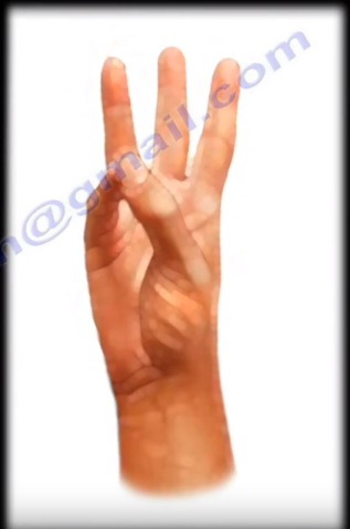 The composite motion involves crossing the thumb over the thumb of the hand toward the base of the little finger. Complete opposition of the thumb may be defined in two ways: by the tip of the thumb touching the base of the fifth digit and by the tip of the thumb touching the tip of the little finger. Therefore, deficiencies in opposition can be quantified by measuring the distance from the tip of the thumb to the base of the little finger or from the tip of the thumb to the tip of the little finger. Alternatively, the degree of opposition may be determined by measuring from the flexor crease of the IP joint to the distal palmar crease over the third metacarpal. A normal span is at least 8 cm. opposition may also be assessed by systematically touching the tip of the thumb to various parts of the hand, a clinical test outlined by Kapandji.
 Position of the hip during the impact decides the injury. In posterior dislocation of the hip, which is the commonest type, the lower limb will be flexed, adducted, and internally rotated. Anterior hip dislocation is rare. It could be a superior anterior hip dislocation. The limb will be extended, abducted, and externally rotated. With an anterior inferior dislocation (obturator type), the extremity will be flexed, abducted, and externally rotated. Hip fractures are different than hip dislocations. Notice that the affected extremity is shortened and externally rotated with a hip fracture. Hip dislocation of any type is an emergency. It must be reduced in less than 6 hours of injury. After reduction of the hip, get a CT scan. Although x-ray is helpful, a CT scan clearly outlines the bony injury. Check the CT scan for congruous reduction, for absence of fracture, and absence of marginal impaction in the acetabulum (with posterior wall fracture, check for marginal impaction). Marginal impaction is more common in posterior acetabular wall fractures and could lead to instability.
  Displaced or comminuted posterior wall fracture could lead to arthritis. Make sure that you have good congruous reduction with no loose bodies or important fractures present. Check for fractures of the acetabulum and the size of the fragment. The size of the posterior wall fracture has an effect on the stability of the hip joint. If congruous reduction of the hip is not obtained, perform open reduction urgently. Open reduction can be done through an anterior approach or a posterior approach. Hip dislocation with or without associated fracture can cause complications. The risk of avascular necrosis depends on the interval between the injury and reduction of the dislocation. Urgent reduction of hip dislocation is mandatory to avoid AVN and interruption of the blood supply which leads to collapse of the femoral head. Reduce the hip and recheck the sciatic nerve function. Always reduce the hip early. Closed reduction should be done in less than 6 hours.
  When injury occurs to the sciatic nerve due to posterior hip dislocation, the common peroneal nerve is usually affected, causing weakness in dorsiflexion of the ankle and loss of toe extension. Injury can occur in varying degrees of severity and it can be missed. Check for foot drop. Movement of the toes may be misleading. Movement of the toes may appear as dorsiflexion, however this really is the result of plantar flexion. Documenting the injury is important to avoid medical legal problems. Injury to the sciatic nerve usually occurs from the dislocation and not from the reduction of the hip. The longer the wait for the reduction of the dislocation, the more the patient is predisposed to sciatic nerve injury. The length of time a hip remains dislocated influences the incidence and the severity of a major sciatic nerve injury. Patient recovery of the sciatic nerve occurs in 60-70% of patients. The patient usually requires an anti-foot drop splint to prevent equinus of the ankle.
  There is approximately 10% incidence of sciatic nerve palsy from posterior hip dislocation. Neurologic examination at the time of injury is usually difficult, however, it is extremely necessary. Check for sensation on the top of the foot. In posterior dislocation of the hip, always look for injuries in the knee such as with a dashboard injury. The force of the injury is usually transmitted from the knee to the hip. There may be an associated posterior cruciate ligament (PCL) injury or a meniscal tear. Examine the knee for injuries and an MRI of the knee may be needed. In cases of high energy trauma, always look at the chest. There might be a tear of the aorta. Check for widening of the mediastinum on chest x-rays. There is concern of deceleration injury involving the aorta. You may apply advanced trauma life support (ATLS) protocol. More flexion, internal rotation, and adduction favors pure dislocation of the hip. Less flexion, internal rotation, and adduction favors fracture dislocation of the hip. Hip dislocation may be associated with acetabular fracture or fracture of the femoral head (Pipkin fracture).
  With Pipkin fracture, as the femoral head dislocates, it hits the posterior wall of the acetabulum and the femoral head fractures. This may be different from an anterior hip dislocation. Anterior hip dislocation will cause impaction of the femoral head or indentation fractures. Classically, Pipkin fracture is a posterior dislocation of the hip and fracture of the femoral head. To treat this, do emergency closed reduction of the hip within 6 hours. Closed reduction is done to avoid avascular necrosis (AVN) of the hip. Reduction of the hip joint and mobilization of the patient with protected weight-bearing crutches for 4-6 weeks. After closed reduction, when the patient has an associated fracture, assess the hip stability, especially if the fragment is not too large. The hip is usually stable if the fragment size of the acetabulum is less than 20%. More than 40%, the hip is unstable. Between 20%-40% fragment size, the hip stability is undetermined. When there is an associated acetabular fracture, the best method to assess the stability of the hip is by examination of the patient under general anesthesia utilizing fluoroscopy. Asses the posterior wall stability with the obturator oblique view. Hip will be in flexion, adduction, and add axial load.
  Check the medial clear space for opening (opening of the medial clear space suggests instability of the posterior wall fracture. Irreducible isolated posterior dislocation; do emergency surgical treatment to reduce the hip. If there is an associated acetabular fracture or femoral head fracture, do urgent closed reduction of the hip dislocation followed by stabilization of either of the fractures if needed according to the protocol. For a posterior hip dislocation with posterior acetabular wall fracture, you must assess the stability of the hip joint by examination under anesthesia after closed reduction. After closed reduction, if the dislocation is not congruent, do open reduction and fixation urgently. For a Pipkin femoral head fracture, do headless screw fixation.
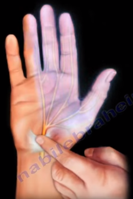 Percussion of the volar wrist crease produces electric sensation distally to the fingers. Phalen’s maneuver is performed by flexing the wrist for 60 seconds. This will increase the carpal tunnel pressure temporarily and produce the symptoms. If the test is positive, the patient will have numbness and tingling in the hand and wrist. The Positive Compression Test (Durkan’s Test) is the most sensitive test. The examiner places even pressure with two thumbs directly over the patients median nerve in the carpal tunnel for about 30 seconds. Reproduction of symptoms in the distribution of the median nerve means that the test is positive for carpal tunnel syndrome. Self-administered hand diagram is extremely helpful (most specific test for carpal tunnel syndrome). The patient should highlight the areas where they are experiencing the symptoms. The patient may complain of thenar atrophy, weakness, or clumsiness of the hand. The patient’s history and examination is an indication for carpal tunnel syndrome.
  Carpal tunnel syndrome is a clinical diagnosis. Carpal tunnel syndrome can be treated by anti-inflammatory medication or activity modification. Activity modification includes avoiding activities that aggravate the symptoms. It can also be treated with neutral wrist splints. It can help night time symptoms because it lowers the carpal tunnel pressure. Functional wrist splints (30o extension) will aggravate carpal tunnel syndrome because it increases the carpal tunnel pressure. At 3 months, 50% of patients will improve with splints. At 18 months, more patients will improve with splints. Sometimes, I use Vitamin B6. There is really no proof that vitamin B6 and physical therapy have any significant effect on improvement of carpal tunnel symptoms. Steroid injections are used for the treatment and for diagnosis of carpal tunnel syndrome if clinical examination or electro-diagnostic test is not clear. If the patient temporarily improves from injection, then the patient will definitely improve from surgery. For steroid injection, mark the intersection of the palmaris longus tendon and the distal palmar crease. Next, go 1 cm proximal and 1 cm ulnar to that site, this will be the point of injection. Use a 25 gauge needle with desired steroid and 1 mL of 1% lidocaine.
  Put the needle at a 45o to the skin of the wrist. Direct the needle towards the base of the thumb and advance the needle distally and slowly. The physician should warn the patient before the injection that if any feeling of numbness, paresthesia, or severe pain exists to let the physician know about it. Injection gives 80% transient improvement and 22% of the patients will be symptom free 1 year after injection. Carpal tunnel release surgery can be open or endoscopic. Surgery is usually done when there is persistence of the symptoms and failure of nonoperative treatment. The injection is a good prognosis for improvement after surgery when the splint no longer works, and when steroid injection only gives temporary improvement (injection is a good prognosis for improvement from surgery). The median nerve is much like a truck passing through a tunnel. The truck (nerve) should be able to pass through the tunnel with ease and without friction. If the tunnel is narrow then the nerve (truck) cannot pass. If you want the nerve to pass, then widen the tunnel. The tunnel is widened by cutting the transverse carpal ligament, as seen in this example. The American Academy of Orthopedic Surgeons (AAOS) recommends doing electrodiagnostic studies before performing carpal tunnel release surgery. Graham stated, that if the patient has a strong history and clinical examination for the diagnosis of carpal tunnel syndrome, then the electrodiagnostic test is unlikely to change the clinical diagnosis. Endoscopic procedure will give a better early rehab.
  The result is the same as with an open release, however incomplete release is a complication of the endoscopic procedure. The pinch strength returns to normal by 6 weeks. The grip strength returns to normal by 12 weeks. At one year, 20% of patients with severe carpal tunnel symptoms will continue to have symptoms. Revision carpal tunnel usually occurs when there is incomplete release. 25% will have no relief. Only 25% will have complete relief. The recurrent motor branch of the median nerve can be injured during the surgery. I want to talk about the anatomy and the variation in distribution of this nerve. After passing through the carpal tunnel, the median nerve gives a branch on the radial side called the recurrent motor branch. The recurrent motor branch is an important nerve supply to the thenar muscles. The recurrent motor branch of the median nerve has multiple variations of the nerve: 30% are subligamentous with recurrent innervation and 20% are transligamentous with recurrent innervation. If this nerve is injured, the patient will get progressive thenar atrophy due to that injury. It is important to cut the transverse carpal ligament far ulnarly to avoid cutting the recurrent motor branch of the median nerve. If you see a patient after carpal tunnel release and that patient has progressive thenar atrophy, this can be explained by the fact that there is an injury to an unrecognized transligamentous motor branch of the median nerve.
 The procedure is referred to as a “salvage” procedure. It is the lesser of two evils or it is the final alternative procedure. This procedure may have a role in cases of displaced femoral neck fractures or in cases of failed internal fixation of femoral neck fractures in debilitated elderly patients. Let’s agree that in the elderly patient, hip prosthesis either unipolar or bipolar and usually cemented, is the ideal surgical procedure for displaced femoral neck fractures, especially if the patient is debilitated and old. In an active elderly patient, total hip replacement should be considered. Sometimes the medical condition and the age of the patient does not support or allow the use of a prosthesis in the elderly. I use the girdlestone procedure in some cases of displaced femoral neck fractures in the elderly, especially if the patient is debilitated and nonambulatory, and when the medical comorbidities are almost prohibitive for surgery. Comorbidities include chronic renal failure, COPD, and congestive heart failure. Even if the prosthesis could be done, the pre-injury cognitive and physical function is predictive of post-operative functional outcome after hip fracture surgery and this select group of patients will not be functional with the prosthesis. The purpose of the Girdlestone procedure is to decrease the pain and to preserve the life of the patient despite the considerable shortening of the extremity. It is an alternative to hospice or alternative care.
  It is the simplest and the least complex procedure for the patient. Counseling to the patient and the patient’s family should be done. The Girdlestone procedure can be done anteriorly or posteriorly. You do not need traction post-operatively. You should get the patient out of bed immediately, and you should do physical therapy early. You will keep the patient in a step down or ICU for a few days after surgery. The patient should be admitted by the geriatric services in cooperation with the trauma services. Surgery should be done within 48 hours or as soon as the patient is optimized medically because that could decrease the mortality rate. Sometimes optimization of the patient is not that easy. The mortality rate is 25% at one year and 6% during hospitalization. The pre-injury mobility is the most significant determining factor for post-operative survival. In patients with femoral neck fractures, surgery done on weekends was associated with an increase in hospital mortality rate, so it is better to do this surgery on week days. If you try to do a simple procedure such as fixation of the displaced femoral neck fracture, the failure rate is about 46% with fixation techniques in the elderly. There is a growing number of people over 90 years of age who will suffer from femoral neck fractures and these patients will need decisions and appropriate care for their situation. Advanced age is associated with increased mortality and poor functional recovery, so we need to think of new ways to approach the increased number of femoral neck fractures in the elderly, and I think that Girdlestone procuedre should be utilized in some select indications.