Despite the sophistication of imaging and the thoroughness of a physician’s physical examination, some injuries remain difficult to diagnose. Obviously, it is imperative for the diagnosis to be made as soon as possible to yield the best possible results for patients. In this second installment of commonly missed orthopaedic injuries, we look at quadriceps tendon rupture, Lisfranc injuries, stress fractures, scaphoid fracures, chance fractures and patient’s with compartment syndrome.

Quadriceps Tendon Rupture (Figure 1)
The quadriceps tendon is located at the top of the patella, with the quadriceps muscle just above the tendon. The quadriceps tendon is essential to moving the knee from a bent to an extended position. As the quadriceps muscle contracts, it pulls the knee’s muscles and tendons including the quadriceps tendon to move the knee into extension. These injuries are sometimes missed, as they are thought to be knee sprains. A combination of physical examination and imaging is needed to diagnose
Lisfranc Injuries (Figure 2)

The Tarso-metatarsal joint complex is referred to as the Lisfranc joint complex. Physicians should look for displacement of the second metatarsal. To confirm the diagnosis, physicians should obtain CT scans or stress views. Swelling in the midfoot region and an inability to weight bear are telling signs of Lisfranc injuries. Surgery is needed for unstable Lisfranc injuries, which is the typical presentation. However, a splint may be used in non-displaced Lisfranc injuries.
Stress Fractures (Figure 3)

Stress fractures are overuse injuries associated with muscle fatigue. Typically, fractures are the result of acute events. Stress fractures, on the other hand, are the result of repetitive stress with a less severe force. Eventually, the fatigued muscle transfers excess stress to the bone causing small cracks in the bone. Physical examination and history are very important factors in the diagnosis of stress fractures as they may not show up on x-rays until several weeks later. While x-rays may show evidence of bone healing, it may be helpful to obtain an MRI or bone scan to confirm the diagnosis. Treatment for stress fractures is usually conservative. Rest is the preferred management.
Scaphoid Fractures (Figure 4)
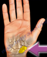
The scaphoid is a small wrist bone located on the thumb side of the hand. Fractures of the scaphoid are sometimes missed, which is potentially problematic as scaphoid fractures have a high risk of delayed union and non-union. In addition, there is a high risk of avascular necrosis with proximal pole fractures. Avascular necrosis is a condition where blood supply to the area of a bone is lost, permanently or temporarily, causing the bone tissue to die. Scaphoid fractures are best evaluated by MRI. Cast immobilization can be utilized for stable and non-displaced fractures, while surgery is needed for displaced fractures.
Chance Fractures (Figure 5)

A Chance fracture is a type of thoracolumbar spine fracture characterized by a horizontal avulsion injury of the vertebral body caused by flexion in which the entire vertebra is pulled apart by a strong tensile force. This type of spine fracture, which is commonly missed during initial evaluation, is also known as a seatbelt injury. Physicians who suspect a Chance fracture should look for associated abdominal injuries such as colon injuries in the pediatric population.
Compartment Syndrome (Figure 6)

Compartment syndrome is an emergent situation which requires immediate attention. It is defined as increased pressure in a closed space that decreases tissue perfusion leading to tissue ischemia and necrosis. Compartment syndrome is often found in the leg and forearm. Five features of compartment syndrome physicians should look for are pain, pallor, paresthesia, pulselessness and paralysis. However, the most important diagnostic sign is pain on passive stretch of the toes and fingers. Compartment syndrome is imperative to diagnose in the impending, because once it is established it may lead to Volkman’s ischemic contracture – the end state of neglected acute compartment syndrome with irreversible muscle necrosis leading to ischemic contracture. Total ischemia of 8 hours produces irreversible changes in muscle. An urgent fasciotomy is the single effective treatment. This is a surgical procedure where the fascia is cut to relieve tension or pressure.
