How To Develop Loyalty In Your Team

The head of the team is responsible for maintaining team morale and leadership. The head of the team is supposed to act as the “glue” that holds the rest of the team together.

loyalty.png

It is important to acknowledge the work that is done by members of your team in order to boost morale. When leadership focuses on building team morale, it creates a positive environment within the office, department or university. It is important to mention the employee by name and show them appreciation for a job well done!

Often the employee does not feel appreciated in their work when they do not receive appreciation from their leaders. Appreciation has the greatest impact on team morale.

Recognition and Appreciation

Allow the employee to feel valued for their continued hard work. Give them praise and publish recognition. Giving recognition to the employee is a positive way to inspire them to use the full extent of their talents and “go the extra mile”. Morale will increase, as well as productivity.

Communication should always occur between the leaders and the employees! Be hands on! Correspondence should not occur simply through email or other non-personal methods. It is important to maintain face to face communication.

Allow the employees the opportunity to lead and receive credit for their accomplishments. As a leader, be there to help and coach others to take responsibility and think for themselves. During times of crisis, lead from the front! During times of comfort, lead from behind!

Engaged Team

An engaged team will be better equipped to achieve the mission and objective of their work. They will believe in what needs to be accomplished with passion and enthusiasm.

loyalty2.PNG

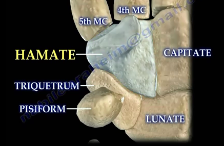
Hamate Fractures

hamate1.PNG

Fractures of the hamate bone are rare, difficult to diagnose, and routine x-rays may not show the fracture. Hamate fractures are classified as either a hook fracture or as a body fracture.

Hamate hook fractures are usually seen in individuals who participate in sports which involve a racquet, baseball bat, or from swinging a golf club.

Swinging of the golf club may cause a hook fracture of the Hamate bone. Missing the fracture can lead to persistent pain from nonunion.

hamate.PNG

Hamate body fractures are associated with axial force trauma, such as a fist striking a hard object, a fall, or from crushing injuries. It may also be accompanied by 4th and 5th metacarpal subluxation. Coronal fractures are the most common type of Hamate body fractures.

There are three types of coronal fractures; Type A (large piece), Type B (moderate piece) and Type C (avulsion). Make sure to watch out for subluxation of the joint due to pull from the Extensor Carpi Ulnaris.

hamate2.png

Hook fractures of the Hamate are best seen by carpal tunnel or supination x-ray views. For hamate fractures, CT scan is the best study. A 30° pronated view is helpful for body fractures.

Clinical Evaluation

Pain will be present, especially with axial loading of the ring and little finger or by grasping an object. The patient will have dismissed grip strength. They may have ulnar and median nerve neuropathy symptoms. The most common findings are pain and tenderness on the ulnar side of the wrist, distal to the wrist joint.

The pull test has been recently described. This is when the palm of the hand is placed into supination, the wrist is in full ulnar deviation, and the fingers of the patient should be flexed. The examiner pulls on the ulnar two digits with the patient resisting the pull. A positive test with pain in the area of the hook indicates a fractures hook of hamate injury. Pain may also be felt due to compression of the ulnar nerve in the Guyon Canal.

Treatment                        

Early immobilization for acute fractures with short arm splint for 6 weeks will be used to avoid a nonunion. For symptomatic nonunion, excision of the fracture fragment will be needed.

Types A and B require open reduction and internal fixation, in addition to stabilization of the joint if needed. Type C requires closed reduction and percutaneous pinning of the fragment for stabilization of the joint. If a closed reduction of the joint is not adequate, open reduction and stabilization of the joint should be done. A displaced fragment with subluxation requires reduction of the joint and stabilization of the joint with K-wires or fixation of the fragment.

When a 4th or 5th carpometacarpal dislocation occurs, one should make special effort to find a coronal fracture of the hamate.


Potential Complications of Bone Fracture

When a bone breaks, the patient may be at immediate risk of acute complications. These include serious vascular or nerve injuries, which occur due to the location of the break, as well as significant blood loss, which can happen if the break injures a major blood vessel. Similarly, a break near a major organ may cause life-threatening distress, such as the compromised respiratory function that occasionally occurs as the result of a rib fracture.

Other immediate risks include fracture blisters, which develop when the surrounding skin attaches to the bone. These can interfere with proper healing and may prompt the development of infection. Physicians must be watchful for these and other injury-generated conditions, including compartment syndromes, which cause pain and potential tissue ischemia.

The most severe complications tend to occur soon after the break, but secondary issues are still possible as the bone heals. These include potentially dangerous infections as well as bone calcifications and complex regional pain syndrome. Bone calcifications primarily require pain management, while regional pain syndrome can be addressed by physiological and psychological therapy. Patients may also be at risk of bone fragments failing to join, a complex condition that may in some cases necessitate surgical intervention.

About the author:

Nabil Ebraheim

Over the course of his career, orthopaedic surgeon Dr. Nabil Ebraheim has conducted extensive research into bone fracture and healing. Dr. Nabil Ebraheim currently serves as the chair of the Department of Orthopaedic Surgery and director of the orthopaedic trauma fellowship at the Medical University of Ohio, where he draws on an in-depth knowledge of fractures and potential complications.

Tension Pneumothorax

Tension Pneumothorax is a life threatening condition in which air leaks outside of the lung and the air is trapped between the pleura and the lung.

tension.png

Air leaks outside of the lung into the pleural cavity. The air is trapped between the pleura and the lung. The air in the pleural space cannot exit! This prevents expansion of the lung and oxygenation. This condition leads to hypoxia and cardiopulmonary collapse. The patient will have acute unilateral chest pain, dysnia, respiratory distress, tachypnea and tachycardia. The patient will also have unilateral decreased or absent breath sounds. As air pushes against the lung, it deviates the trachea to the other side.

tension2.png

Treatment

Treatment will include needle decompression. Insert the needle at the 2nd intercostal space, mid clavicular line, followed by the insertion of a chest tube.